Fecha de publicación: junio 28, 2018| Publicado en el Número 3

Caracterización molecular de la coexistencia de haploinsuficiencia 18q y duplicación 18p en paciente con fenotipo sindrómico complejo

Fátima Gimeno-Ferrer1, David Albuquerque1,2, María Capataz Ledesma3, Carolina Monzó1, Guillermo Gervasini4, Francisco Barros Angueira5, José María Carbonell6, Goitzane Marcaida Benito1, Raquel Rodríguez-López1, Enrique Galán Gómez3,7*

1 Grupo de Genómica, Fundación de Investigación Consorcio Hospital General Universitario de Valencia, Avenida Tres Cruces 2 46014 Valencia, España.

2 Centro de Investigación de Antropología y Salud (CIAS), Departamento de Ciencias de la Vida, Universidad de Coimbra, Calçada Martim de Freitas 3000-456 Coimbra, Portugal.

3 Unidad de Genética Pediátrica, Hospital Materno Infantil, Av. Damián Tellez Lafuente s/n 06010 Badajoz, España.

4 Departamento de Medicina y Cirugía Terapéutica, Facultad de Medicina, Universidad de Extremadura, Av. Elvas s/n 06071 Badajoz, España.

5 Fundación Pública Gallega de Medicina Genómica, CIBERER, Grupo de Medicina Genómica, Hospital Clínico, Santiago de Compostela, Universidad de Santiago de Compostela, Praza do Obradoiro s/n 15782 Santiago de Compostela, España.

6 Servicio de Inmunología del Hospital Infanta Cristina, Av. Elvas s/n 06071, Badajoz, España.

7 Departamento de Ciencias Biomédicas. Terapéutica, Facultad de Medicina, Universidad de Extremadura, Av. Elvas s/n 06071 Badajoz, España.

Resumen

Desde que los arrays de SNPs/CNVs de genoma completo se han erigido como la principal estrategia diagnóstica en la práctica clínica en el ámbito de la neuropediatría, se han identificado numerosos desequilibrios cromosómicos causales de discapacidad intelectual considerada idiopática previamente. Su uso ha favorecido la descripción exhaustiva de las bases moleculares de numerosos nuevos síndromes, con especial eficiencia en aquellos asociados a múltiples anomalías congénitas y/o rasgos dismórficos. Para describir exhaustivamente la correlación fenotipo-genotipo en una paciente de 2.5 años cuyo cariotipo evidenció una alteración citogenética compleja con dos líneas en mosaico, se hibridó su ADN genómico en un microarray de alta densidad de SNPs/CNVs. El resultado del microarray detectó una duplicación de 12.5 Mb de la región cromosómica 18p11.32p11.21 (chr18:12602631_telómero) y una deleción de 20.3 Mb de la región cromosómica 18q21.32q23 (chr18:57691236_telómero). El cariotipo reveló la existencia de una línea celular con porcentaje del 92% portadora de la duplicación y de la deleción, y una segunda línea celular minoritaria (8%) que presentaba un cromosoma en anillo portador de la deleción detectada en el brazo q. La caracterización molecular de las regiones cromosómicas duplicadas y en haploinsuficiencia, identificaron las alteraciones causales de los principales signos y síntomas aparecidos en la paciente. Aunque el cariotipo convencional se considera actualmente una técnica diagnóstica de apoyo o segunda línea, fue clave para establecer la etiología de la alteración que portaba la paciente. En este caso permitió la interpretación correcta del resultado del array y sólo con ambas pruebas se pudo concluir la existencia del cromosoma 18 en anillo, así como el porcentaje exacto de mosaicismo de cada línea. La capacidad deletérea de los genes OMIM dominantes implicados, permitió el seguimiento clínico riguroso de la paciente y la descripción del fenotipo observado. La inclusión de tal información en las bases de datos específicas, junto a los resultados moleculares obtenidos del array, permite su comparación con alteraciones genéticas detectadas en otros pacientes y cuyas localizaciones cromosómicas sean solapantes.

Palabras clave: haploinsuficiencia 18q, duplicación 18p, SNP/CNV array, mosaicismo, fenotipo sindrómico

Molecular characterization of the coexistence of 18q haploinsufficiency and 18p duplication, causal of a complex syndromic phenotype

Since genomic SNPs/CNVs arrays were implemented as a diagnostic tool in clinical settings to search for the cause of idiopathic intellectual disability, chromosomal imbalances have been precisely described as being the cause of many new syndromes, especially when they are associated with multiple congenital anomalies and/or dimorphism. High-density SNPs/CNVs microarray was used to delineate genotype-phenotype correlation in a 2.5 year-old girl who carried a mosaic characterized by a predominant cell line representing 92% which carried a duplication of 12.5 Mb of the 18p11.32p11.21 chromosomal region (chr18:12602631_telomeric) and a deletion and a deletion of 20.3 Mb of the 18q21.32q23 chromosomal region (chr18:57691236_telomeric) and a second minor cell line (8%) presented a ring chromosome, carrying the deletion of 20.3 Mb of the 18q21.32q23 chromosomal region. The microarray analysis identified the genetic causes for the specific phenotype of the patient, whose more evident signs and symptoms were mainly associated to the genomic regions duplicated and in haploinsufficiency. Although the conventional karyotype is currently considered a diagnostic technique of support or second line, it was key to establish the etiology of the alteration that the patient carried. Its performance allowed the correct interpretation of the result of the array, whose characterization facilitated its correlation with the phenotypic features of the patient. From both tests, it was possible to conclude the existence of ring chromosome 18, as well as the percentage of the mosaic lines. The deleterious capabilities of the affected OMIM and dominant genes were evaluated along her period of life. A rigorous clinical following up in that patient included valorous phenotypic data to the array data bases, and will make less difficult to hypothesize about prognosis in other individuals who carry overlapping similar high risk genetic alterations.


Keywords: 18q haploinsufficiency, 18p duplication, SNP/CNV array, complex phenotype

Contenido del artículo

INTRODUCCIÓN

La implementación de las técnicas de hibridación de genoma completo ha optimizado muy notablemente la eficiencia diagnóstica y detección de anomalías cromosómicas, detectando y caracterizando las mismas y superando ampliamente la eficiencia del cariotipo convencional (Dhillon 2014). Actualmente, se acepta la utilidad clínica de esta tecnología de arrays de polimorfismos de nucleótido único y variantes en número de copias (SNPs/CNVs) como base del algoritmo diagnóstico genético para los pacientes afectados de discapacidad intelectual, así como del protocolo para el cribado de pacientes que además han desarrollado fenotipos sindrómicos. Los resultados

obtenidos por este tipo de matrices definen exhaustivamente las alteraciones citogenéticas detectadas por técnicas convencionales, pero sobre todo de un gran número de nuevas pérdidas y ganancias cromosómicas crípticas implicadas en este grupo de patologías. El aumento progresivo de la cobertura de sus diseños, ha ido permitiendo la caracterización de nuevas anomalías cromosómicas subcrípticas, y su posterior correlación fenotípica con otros cuadros clínicos sin etiología conocida. La consecuencia inmediata de la aplicación rutinaria de esta tecnología, ha señalado una serie de nuevos genes de susceptibilidad candidatos a ser valorados como bases moleculares causales del desarrollo de discapacidad mental y/o rasgos dismórficos.

La descripción de los signos y síntomas clínicos observados en los pacientes con fenotipos extremos, se realiza actualmente de forma homogénea en base a un esfuerzo ingente realizado para denominar cada uno de ellos según la base de datos Human Phenotype Ontology (HPO); cada uno de los pacientes afectados exhibe un amplio y complejo conjunto propio de signos y síntomas clínicos, debido al desarrollo individual de distintas anomalías en órganos y rasgos físicos. En relación con esto, a pesar de que los arrays de SNPs/CNVs se aplican homogéneamente como herramienta diagnóstica esencial de búsqueda de la causa de discapacidad intelectual idiopática en niños (Shaffer 2007; D’Angelo 2012), la caracterización fenotípica sigue siendo aún somera y difícil de catalogar. La descripción de variantes causales progresa exponencialmente, constituyendo una valiosísima base de datos que detalla la enorme variedad de desequilibrios cromosómicos nuevos y únicos descritos. El valor de su información se basa en su aplicabilidad al ámbito clínico y su asociación definitiva con alteraciones clínicas no filiadas previamente.

En este artículo presentamos el caso clínico de una niña con un conjunto complejo de signos y síntomas, entre los que destaca un evidente déficit del crecimiento, asociados fundamentalmente a la haploinsuficiencia de los genes OMIM: CTDP1, TNFRSF11A, MC4R, PIGN, CDH19, TMX3, RTTN, ZNF407, TSHZ1, MBP y SALL3.

Para el diagnóstico de la paciente se consideró el uso del array de hibridación genómica como método de primera elección, para la descripción rápida y precisa de las posibles anomalías cromosómicas existentes. Los resultados obtenidos definieron la correlación del genotipo con el fenotipo observado. Sin embargo, el uso de cariotipo convencional fue crucial para entender la complejidad de la alteración que portaba la paciente, así como la etiopatogenia de los grandes reordenamientos citogenéticos y mosaicismos que presentaba. Fue de especial interés la detección y descripción citogenética de parte de la alteración existente en la línea celular portadora del cromosoma 18 en anillo, cuyo porcentaje fue calculado menor al 10%.

DESCRIPCIÓN DEL CASO CLÍNICO

Se presenta el caso de una niña de 6 meses que fue remitida para asesoramiento clínico y posterior tratamiento, siendo la segunda hija de padres no emparentados, jóvenes y sanos. No existían antecedentes familiares destacables. El embarazo fue controlado, terminando con un parto vaginal a las 36+6 semanas de gestación. El peso de la paciente al nacer fue 2.240 g (P10), una talla de 44 cm (P10-25) y un perímetro craneal de 31 cm. La puntuación Apgar fue de 9/9, al minuto y a los cinco minutos respectivamente. En el periodo neonatal, la paciente presentó taquipnea transitoria e ictericia neonatal no inmune que requirió fototerapia de menos de 24 horas. Durante la hospitalización, sufrió una infección por estafilococo y epidermitis séptica, tratada con antibióticos intravenosos sin complicaciones.

Los exámenes físicos de la paciente mostraron hipotonía axial moderada y rasgos dismórficos como hipertelorismo, puente nasal plano, orejas displásicas y pliegues de piel redundantes. A nivel abdominal, presentaba una hernia umbilical leve. La paciente padecía una malformación anorectal con una fístula perineal con abertura anal ectópica con recorrido en horquilla. Mostraba abducciones irreducibles en miembros inferiores y pies planos severos. Se detectó la cavidad septum pellucidum y cavum vergae mediante ecografía. Las emisiones otoacústicas y exploración auditiva del tronco encefálico mostraron potenciales patológicos con déficit de conducción auditiva. La TAC del peñasco del temporal mostró estenosis de ambos conductos auditivos. Se le diagnosticó mediante ecocardiografía un defecto del cierre del septo atrial de tipo ostium secundum y un ductus arterioso persistente.

La paciente llevó audífonos desde los 5 años y vendajes en miembros inferiores, con un programa intenso de fisioterapia. Sufrió un episodio de bronquiolitis leve a los 8 meses y varios episodios de broncoespasmos leves-moderados, con ingreso hospitalario a los 11 y 12 meses de edad. Presentaba un evidente retraso psicomotor (consiguió sostener la cabeza a los 8 meses, mantenerse sentada a los 17 meses y levantarse a los 3 años), retraso en el lenguaje (sólo balbuceaba) e hipotonía. Su estudio cerebral mediante resonancia magnética mostró hipoplasia del cuerpo calloso, hipertrofia de la masa intermedia del tálamo y escasa diferenciación entre la materia gris y blanca. El electroencefalograma fue normal.

Desde los 9 meses la talla baja era manifiesta, midiendo 80 cm a los 2 años (en P1, correspondiendo a -2.35 DE) con un peso por debajo del P1 (9 kg, correspondiente a -2.52 DE). Tenía una microcefalia por debajo del P1 desde el nacimiento (-3.64 DE) con plagiocefalia con asimetría posicional y una asimetría de la cara con menor desarrollo de la parte izquierda. Presentaba además fisuras palpebrales horizontales, ojos pequeños con epicanto antimongoloide, estrabismo en el ojo izquierdo, raíz nasal amplia y deprimida, nariz pequeña en punta y boca pequeña con paladar alto. Presentó micrognatia leve y orejas de implantación baja. El examen del tórax era normal, aunque con incremento de la distancia entre mamilas; el abdomen, la cintura pélvica y las caderas eran normales.

La última evaluación por su pediatra especialista en dismorfología fue a los 4.6 años y mostró mejoría general. Aunque la mayoría de los rasgos dismórficos persistieron, se detectaron cambios en el fenotipo de la paciente como la normocefalia y la mejora de la plagiocefalia asimétrica posicional, ojos normales con epicanto bilateral y mejora de su estrabismo. Entonces se describió la existencia de un surco subnasal corto.

ESTUDIOS CITOGENÉTICOS Y MOLECULARES

Después de obtener el consentimiento informado, se realizó un cultivo de linfocitos de sangre periférica de la paciente y sus progenitores para llevar a cabo el cariotipo de bandas GTG de alta resolución (Figura 1). La alteración cromosómica fue molecularmente caracterizada usando array citogenético de genoma completo 2.7 M de Affymetrix (Affymetrix Inc., Santa Clara, CA, USA) (Figura 1). Los datos fueron recolectados usando Gene Chip Scanner 3000 Dx y los ficheros CEL fueron analizados con el software Chromosome Analysis Suite (ChAS v1.1, Affymetrix inc, Santa Clara, CA, USA). La referencia para la comparativa de datos obtenidos fue hg19. Las CNVs detectadas en la paciente fueron comparadas con la Database of Genomic Variants (DGV; http://projects.tcag.ca/variation) y con el International Standards for Cytogenomic Arrays Consortium (Public ISCA database; https://www.iscaconsortium.org). La Database of Chromosomal Imbalance and Phenotype in Humans (DECIPHER; https://decipher.sanger.ac.uk) fue usada como referencia principal para la evaluación de la posible capacidad deletérea de las alteraciones detectadas.

Se realizó análisis por secuenciación Sanger automática bidireccional de toda la región exónica y secuencias flanqueantes del gen MC4R, a partir de ADN genómico en la paciente. Las secuencias de los cebadores usados para la amplificación fueron diseñadas con el software Primer 3 (Untergasser 2012) (secuencias disponibles bajo solicitud). La secuenciación se realizó en secuenciador ABI 3130 DNA Analyzer (Applied Biosystems, Foster City, CA, USA) usando Big Dye Terminator v3.1 Cycle Sequencing Chemistry (Applied Byosystems, Foster City, CA, USA), de acuerdo con el protocolo recomendado por el fabricante. La asignación de bases fue realizada con el programa Sequencing Analysis v5.2 (Applied Biosystems, Foster City, CA, USA). Las secuencias obtenidas fueron analizadas con los softwares Staden package (Staden 1996) y SeqScape v2.5 (Applied Biosystems, Foster City, CA, USA) y comparadas con la secuencia de referencia (ENSG00000166603).

RESULTADOS

El cariotipo convencional de alta resolución y bandeo GTG evidenciaron que la paciente presentaba dos líneas celulares en mosaico: el 92% de las células portaban un cromosoma 18 anormal con una duplicación de la región 18p y una deleción de la región 18q, y el 8% de las células portaban un cromosoma 18 en anillo. Estas alteraciones cromosómicas fueron molecularmente caracterizadas por el array. La fórmula del cariotipo fue mos 46,XX,der(18)add(18)(q21.3)del(18)(q21.3)dn[46]/46,XX,r(18)(p?q21.32)dn[4], siguiendo la normativa establecida en la International System for human Cytogenetic Nomenclature (ISCN). Por tanto, la duplicación 18p del cromosoma 18 derivado se localiza en 18q21.3 tras sufrir la deleción. El estudio citogenético convencional de ambos padres mostró en ambos un cariotipo normal.

El análisis del array detectó tanto la duplicación de 12.5 Mb en el brazo corto (de chr18:12602631 hasta el telómero) del cromosoma 18 derivado que portaba la línea celular mayoritaria, así como la deleción de 20.3 Mb en el brazo largo (de chr18:57691236 hasta el telómero) observada tanto en la línea mayoritaria (92% células con cromosoma 18 derivado) como en las células portadoras del cromosoma en anillo (calculado en un 8%). La fórmula molecular obtenida con el array de genoma completo fue arr[GRCh37]18p11.32p11.21(chr18:136226_12602631)x3, 18q21.32q23(chr18:57691236_78014123)x1, usando la nomenclatura del ISCN (Figura 1). El resto de los resultados obtenidos fueron normales.


haploinsuficiencia 18q
Figura 1. Caracterización molecular de la paciente por array de SNPs/CNVs y cariotipo convencional. A) Imagen obtenida del programa ChAS mostrando las regiones incluidas en la deleción (barra en rojo) y en la duplicación (barra en azul), ambas portadas por la paciente. La fórmula molecular del microarray, arr[GRCh37]18p11.32p11.21(chr18:136226_12602631)x3,18q21.32q23(chr18:57691236_78014123)x1dn, representa la duplicación de 12.5 Mb 18p subtelomérica y la deleción de 20.5 Mb 18q subtelomérica. B) Representación de los genes OMIM delecionados (panel superior) y duplicados (panel inferior) (NC _000018.10 grCh38). C) Cariotipo convencional de alta resolución de la paciente con la fórmula citogenética mos 46,XX,der(18)add(18)(q21.3)del(18)(q21.3)dn[46]/46,XX,r(18)(p?q21.32)dn[4], evidenciando dos líneas en mosaico: 92% de células con un cromosoma 18 anormal con una duplicación del brazo p en la región 18q21.3 y la deleción del brazo q y 8% con un cromosoma 18 en anillo. El cromosoma en anillo era portador de la deleción en el brazo q y una deleción telomérica en el brazo p. Esta última no fue detectada en el array, debido a su bajo porcentaje y el solapamiento con la duplicación de la línea mayoritaria. Los cromosomas normales están a la izquierda y las copias reordenadas están señaladas por flechas a la derecha.

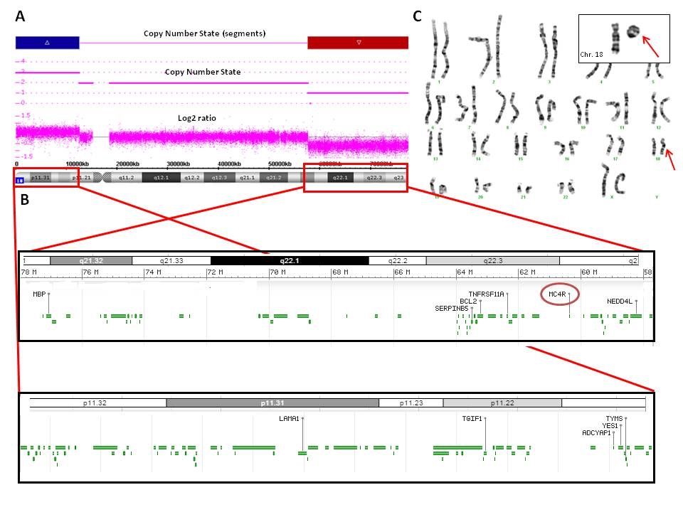
La zona duplicada afectaba 12.5 Mb, que incluía un total de 53 genes. Destacaban los genes con entrada en la base de datos OMIM: NDUFV2, LPIN2, USP14, SMCHD1, TGIF1, APCDD1, GNAL, AFG3L2 y IMPA2.

La deleción mostraba un tamaño de 20.3 Mb y punto de rotura proximal en la posición 57,691,236 del brazo largo del cromosoma 18 (ChAS software (hg19)).  En dicha área delecionada se localizaban un total de 32 genes, de los cuales CTDP1, TNFRSF11A, MC4R, PIGN, CDH19, TMX3, RTTN, ZNF407, TSHZ1, MBP y SALL3 tenían entrada en la base de datos OMIM (Figura 1).

Los resultados del array no revelaron datos acerca de la caracterización molecular de otra pequeña deleción distal en 18p, que portaba el cromosoma 18 en anillo. Dicha deleción distal 18p, responsable de originar el cromosoma en anillo, pudo no ser detectada por el array fundamentalmente por su coincidencia con la duplicación de la línea mayoritaria. El  porcentaje calculado para esta línea celular (8%), no fue insuficiente para detectar la deleción localizada a nivel del brazo q.

Las secuencias del gen MC4R, cuya alteración está implicada en el desarrollo de obesidad, fueron normales, sin mutaciones conocidas ni otras variantes no descritas. Otros genes como CTDP1, TNFRSF11A, PIGN, CDH19, TMX3, RTTN, ZNF407, TSHZ1, MBP y SALL3 no se consideraron candidatos a ser secuenciados porque su capacidad funcional individual en humanos no está bien caracterizada.

DISCUSIÓN

Presentamos el caso de una niña que mostraba un conjunto de signos y síntomas que resultan en un fenotipo complejo, molecularmente caracterizado por un cromosoma 18 derivado portador de una duplicación de 18p11.32p11.21 (en el 92% de las células) y una deleción en 18q21.32q23, también presente en la línea celular en anillo (en el 8% de las células), que causaba fundamentalmente la haploinsuficiencia conjunta de los genes CTDP1, TNFRSF11A, MC4R, PIGN, CDH19, TMX3, RTTN, ZNF407, TSHZ1, MBP y SALL3. La caracterización de la deleción de novo de 20.3 Mb (arr[GRCh37]18q21.32q23(chr18:57691236_78014123)x1) evaluó además la capacidad deletérea del conjunto de 32 genes contiguos delecionados. El último examen clínico realizado a los 4.6 años confirmó la mayoría de los signos, síntomas y malformaciones aparecidas desde el nacimiento, correlacionándolos con los resultados obtenidos con el microarray.

La deleción 18q fue comparada con otras deleciones similares descritas en otros pacientes, las cuales también incluían el gen MC4R, (Hale 2000; Cody 2009; Margarit 2012; Versacci 2005), permitiendo la correlación genotipo-fenotipo en dicha serie y en nuestra paciente. Los genes delecionados en esta región (de 18q21.32 a 18q23) habían sido previamente asociados fundamentalmente con microcefalia, atresia del canal auditivo y defectos de la mielinización (Hale 2000; Margarit 2012). El retraso psicomotor y de lenguaje, así como el déficit neurológico se ha relacionado también con pérdidas similares de estos genes contiguos (Cody 2007). El gen TSHZ1 está asociado con la atresia auditiva congénita, con ausencia bilateral o formación incompleta del canal auditivo, y también se relaciona con alteraciones en el oído medio (Feenstra 2011). Estos signos se han detallado en aproximadamente el 66% de los pacientes con deleción terminal en 18q (Veltman 2003).

Aunque la obesidad es una patología compleja cuya etiología genética se ha relacionado con un amplio número de genes, se han descrito la haploinsuficiencia (Cody 1999) y el déficit función de MC4R (Abdullah 2016; Farooqi 2005; Turner 2015) como causas monogénicas no sindrómicas mayoritarias. Se conoce muy poco acerca del comportamiento de las alteraciones de los genes de susceptibilidad a obesidad no sindrómica, asociados a desequilibrios cromosómicos; las series analizadas tratan de identificar en los pacientes con discapacidad intelectual asociada a obesidad, unas bases moleculares diferentes (D’Angelo 2012).

No se observaron los efectos esperados por la deleción del gen MC4R en nuestra paciente, quizás por estar contrarrestados por la gran capacidad deletérea de las alteraciones que portaba y que causaban su grave fenotipo sindrómico. Se analizaron en la paciente parámetros específicos para detectar evolución del crecimiento, signos y síntomas de fenotipo metabólico y/o daño neurocomportamental por la haploinsuficiencia del gen MC4R. Sin embargo, la evaluación en la paciente fue normal. Adicionalmente, la secuenciación automática Sanger de este gen confirmó que la paciente no portaba ninguna mutación, mostrando un patrón molecular normal en la única copia del gen que portaba. Por tanto, el no desarrollo de obesidad en la paciente podría deberse al complejo genotipo, compensando la haploinsuficiencia del gen MC4R.

El retraso en el desarrollo fue siempre evidente en la paciente. Entre los pacientes portadores de deleciones terminales 18q desde la banda q21 hacia el telómero, suele describirse una escasa ganancia de peso y el déficit en el crecimiento físico después del nacimiento. Se atribuyen a la hipotonía, al reflujo gastroesofágico y paladar ojival, los cuales pueden ocasionar graves dificultades para succionar, tragar y/o engancharse en el pecho (Hale 2000; Feenstra 2007) (http://www.rarechromo.org/information/Chromosome%2018/18q%20deletions%20from%2018q21%20and%20beyond%20FTNW.pdf). Sin embargo, un porcentaje de los pacientes que llegaron a edad adulta, alcanzaron pesos normales e incluso desarrollaron sobrepeso; algunos autores sí han sido descrito la aparición de obesidad en un número destacable de individuos portadores de 18q- (Feenstra 2007).

Esta anomalía cromosómica fue aún más compleja ya que la paciente además presentaba en el cromosoma 18 derivado (mosaico del 92% de las células),  una duplicación de 12.5 Mb del telómero del brazo corto (trisomía) del cromosoma 18, localizada en 18q21.3, y la deleción 18q. La duplicación 18p es una anomalía cromosómica rara. Hay pocos casos descritos en la literatura (Mabboux 2007). La mayoría de pacientes tienen un fenotipo aparentemente normal con pocos rasgos dismórficos y puede manifestarse con discapacidad intelectual de grado variable. La paciente presentó una malformación anorectal consistente en una fístula perineal con ano ectópico y abierto. Se han descrito casos de pacientes portadores de trisomía parcial de la región 18p-q12 y que padecían malformaciones anorectales, sin definirse la región causal implicada (Schramm 2011).

Del conjunto de genes localizados en la duplicación, destaca IMPA2 por su asociación a mayor susceptibilidad a padecer convulsiones febriles (Nakayama 2004).

Parece esencial en este caso la aceptación de que la especie humana tolera mejor el exceso de material genético que su pérdida. Nuestra paciente muestra unos hallazgos clínicos fundamentalmente asociados con la pérdida de material 18q. Sin embargo, la malformación anorectal puede estar más relacionada con la duplicación 18p (Schramm 2011). Como en la mayoría de los pacientes portadores de estas alteraciones complejas, ambos progenitores no presentaron problemas clínicos ni de desarrollo y mostraron tener cariotipo normal; la alteración citogenética compleja fue considerada de novo en la paciente.

Aunque la hibridación de ADN genómico mediante array de alta resolución tiene un papel principal en el diagnóstico de la discapacidad intelectual, el cariotipo convencional fue clave en este caso para identificar el mosaicismo, las reorganizaciones cromosómicas y el cromosoma en anillo. Por tanto, es necesario realizar esta técnica convencional para confirmar genotipos citogenéticamente complejos como el que aquí se expone y postular el mecanismo molecular de origen.

Resumimos cómo la línea celular predominante formada por un cromosoma 18 derivado, que representa el 92% del mosaico, apareció debido a un evento de duplicación de la región 18p localizada en la región 18q21.3, la cual se ha observado y se caracterizado con el cariotipo convencional y el array de genoma completo respectivamente. Este cromosoma derivado además presenta una la deleción en 18q. La línea celular minoritaria, que representa el 8% del mosaico, apareció por dos eventos de deleción, uno en la región 18q distal, el cual es una deleción de gran tamaño caracterizada por el array y también presente en la línea mayoritaria, y una deleción pequeña en la región 18p no detectada por el array debido al porcentaje pequeño de la línea celular así como a la presencia de la duplicación en la línea celular mayoritaria, enmascarando la pequeña deleción responsable del cromosoma en anillo.

 CONCLUSIÓN

Utilizando la técnica del microarray de SNPs/CNVs, se ha descrito el fenotipo complejo de una paciente portadora de dos líneas celulares en mosaico, siendo la mayoritaria la que presenta un cromosoma 18 derivado con una duplicación 18p y una deleción 18q y siendo la línea minoritaria la que presentó un cromosoma en anillo originado por una gran deleción 18q y una pequeña deleción en la región 18p distal no detectada por el array. En el caso de fenotipos complejos, el cariotipo convencional es esencial para resolver el diagnóstico molecular. La evaluación de los genes OMIM incluidos en la deleción mostró que la haploinsuficiencia 18q correlaciona con la mayoría de signos y síntomas del fenotipo de la paciente. Además, la haploinsuficiencia del gen MC4R no alteró el metabolismo ni se asoció con el desarrollo de obesidad en la paciente. La descripción del genotipo y de las características fenotípicas de la paciente facilitará la evaluación de otros pacientes portadores de alteraciones similares.

CONFLICTO DE INTERESES

Todos los autores reconocen y revelan no tener conflicto de interés que declarar. Este trabajo fue apoyado por ISCIII, Madrid con apoyo financiero (proyectos 07PI594, 10PI357). David Albuquerque es beneficiario de una beca postdoctoral (SFRH/BPD/109043/2015) de Fundação para a Ciência e a Tecnologia (Portugal).

AGRADECIMIENTOS

Agradecemos el conocimiento de Ines Quintela, la persona al cargo de la plataforma de Affymetrix en CeGen, Nodo de Santiago de Compostela, ubicado en el Hospital Clínicos Universitario, dirigido por el Dr. Ángel Carracedo.

INTRODUCTION

Last years, the development of microarray technology has improved the diagnosis and detection rate of chromosomal abnormalities over the conventional karyotype (Dhillon 2014). Clinical efficiency of the single-nucleotide polymorphism/copy number variation (SNP/CNV) array technology has been widely established. In our days, it constitutes the base of an efficient genetic diagnostic algorithm to intellectual disability, as a screening protocol in patients with syndromic phenotype. The obtained results describe the prevalence of the genetic etiologies in patients with syndromic intellectual disability of unknown etiology and detail the variability of cryptic chromosomal imbalances implicated. Characterizations of subcryptic chromosomal anomalies are also being produced, and the exhaustive phenotypic correlation description of the new syndromes is progressively considered essential. New candidate susceptibility genes are appearing implicated in the development of dimorphic mental incapacity.

The description of the early-onset intellectual disability associated to the development of an extreme phenotype, allowed to detail the genotype-phenotype correlation focused on the clinical signs and symptoms apart from intellectual disability, as organ-specific developmental abnormalities and dimorphic traits description. In this regard, although SNP/CNV arrays have been implemented as a diagnostic tool to look for the cause of idiopathic intellectual disability in children (Shaffer 2007; D’Angelo 2012), the use of these arrays has not yet been rigorously used for the exhaustive characterization of the appearing syndromic phenotype. After improving the phenotypic data about analyzed patients, a variety of chromosomal imbalances have been accepted as the cause of many unaffiliated clinical alterations.

We report here a clinical case of a girl with a rare complex set of signs and symptoms, including deficient growth measures and haploinsufficiency for the OMIM genes CTDP1, TNFRSF11A, MC4R, PIGN, CDH19, TMX3, RTTN, ZNF407, TSHZ1, MBP and   SALL3.

We implemented array analysis as a first method of choice for a fast and accurate detection of chromosomal abnormalities in this patient and for a better phenotype/genotype characterization. However, the use of conventional karyotype was crucial to understand better this correlation and to observe big rearrangements such as the ring chromosome 18 presented in mosaic in the patient.

CLINICAL REPORT

A 6-month-old girl was referred for clinical assessment and treatment. She was the second daughter of unrelated young and healthy parents.  Pregnancy was well tolerated and controlled. The normal vaginal delivery was at 36+6 weeks of gestation. Birth weight was 2.240 g (P10); length 44 cm (P10-25) and head circumference 31 cm. Apgar score were 9/9 at one and five minutes respectively. In the neonatal period she presented transient tachypnea and non-immune neonatal jaundice that required phototherapy for less than 24 hours. During hospitalization, she suffered a staph and epidermidis sepsis, which was treated with intravenous antibiotics without complications.

Physical examinations showed moderate axial hypotonia and highlighted many dimorphic features such as hypertelorism, flat nasal bridge, dysplastic ears, and redundant skin fold. At abdominal level a minimal umbilical hernia was noted. The patient had an anorectal malformation consistent in a perineal fistula, with an ectopic anal opening in backplane fourchette. At locomotor level, she had irreducible abducts and severe valgus feet. Cerebral ultrasound detected the cavity of septum pellucidum and vergae. The otoacoustic emissions and brainstem auditory evoked potential pathology, showing a conductive hearing loss. The temporary rock TAC showed stenosis of both ear canals. An echocardiogram was performed and an atrial septal defect, ostium secundum type and patent ductus arteriosus were diagnosed.

The patient wore hearing aids from 5 months of age due to conductive hearing loss. Flat feet valgus was resolved through bandages and physiotherapy. She had a mild bronchiolitis episode at 8 months old and has had several mild-moderate episodes of bronchospasm. She was admitted at hospital twice with 11 and 12 months of age. She had psychomotor delay (she held her head up at the age of 8 months, sat up at 17 months and could get up at 3 years), language delay (she only babbles) and was hypotonic. A brain magnetic resonance imaging (MRI) showed hypoplasia of the corpus callosum, intermediate mass thalamus hypertrophy and poor differentiation between gray and white matter. The electroencephalogram was normal.

The girl showed growth delay, at the age of 2 years her weight was of 9kg (below P1, corresponding to -2.52 SD) and her size presented a fall from 9 months of age, having a height of 80 cm at 2 years (in p1, corresponding to -2.35 SD). Microcephaly was appreciated, remaining the percentiles from birth below p1 (-3.64 SD).  The cardiological examination was normal after Inter-Auricular Communication (CIA) and dose-area product (DAP). The dismorphologic examination at 2.5 years showed: microcephaly, asymmetric positional plagiocephaly and less development of left hemiface. She continued using headphones. There were horizontal palpebral fissures, small eyes with bilateral epicanthus, strabismus on the left eye, depressed and broad nasal root, small nasal tip and small mouth with high palate. She had mild micrognathia and low-set ears. There was normal chest with increased internipples distance and normal abdomen. The anus was very small and near to fourchette. Limbs were normal.

The last dismorphologic examination at 4.6 years showed better general and global state. Most dismorphic traits persevered. Changes in the patient’s phenotype included normocephaly, improvement of asymmetric positional plagiocephaly, normal eyes with bilateral epicanthus and mild strabismus. In addition, short philtrum was described.

CYTOGENETIC AND MOLECULAR STUDIES

After obtaining informed consent, peripheral blood lymphocyte cultures of the patient and the parents were set up by standard techniques for karyotyping with high resolution GTG banding (Figure 1). Chromosomal alteration was molecularly characterized using the Affymetrix Cytogenetics Whole-Genome 2.7 M Array (Affymetrix Inc., Santa Clara, CA, USA) (Figure 1). Data was collected using Gene Chip Scanner 3000 Dx and CEL files were analyzed using Chromosome Analysis Suite software (ChAS v1.1, Affymetrix Inc, Santa Clara, CA, USA). The annotation file used was hg19. Detected CNVs were compared with the Database of Genomic Variants (DGV; http://projects.tcag.ca/variation) and with the International Standards for Cytogenomic Arrays Consortium (Public ISCA database; https://www.iscaconsortium.org). The Database of Chromosomal Imbalance and Phenotype in Humans (DECIPHER; https://decipher.sanger.ac.uk) was used as main resource for evaluating the clinical significance of the detected alterations.

Bidirectional sequence analysis of genomic DNA including the whole exonic region of the MC4R gene was performed on the patient. Primer sequences used for amplification were designed with Primer 3 software (Untergasser 2012) (primer sequences available upon request). Sequencing was performed on an ABI 3130 DNA Analyzer (Applied Biosystems, Foster City, CA, USA) using Big Dye Terminator v3.1 Cycle Sequencing Chemistry (Applied Biosystems, Foster City, CA, USA), according to protocols recommended by the manufacturer. Base calling was performed with Sequencing Analysis v5.2 (Applied Biosystems, Foster City, CA, USA). The obtained sequences were analyzed with the Staden package software (Staden, 1996) and the SeqScape Software v2.5 (Applied Biosystems, Foster City, CA, USA), and compared with the normal sequence (ENSG00000166603).

RESULTS

High-resolution karyotype by standard and GTG banding techniques evidenced that the patient had two mosaic cell lines: 92% of the cells had an abnormal chromosome 18 with a 18p duplication and 18q deletion and 8% had a ring chromosome 18. Karyotyping revealed was mos 46,XX,der(18)add(18)(q21.3)del(18)(q21.3)dn[46]/46,XX,r(18)(p?q21.32)dn[4], following the International System for Human Cytogenetic Nomenclature (ISCN). Therefore, the 18p duplication of the derivative chromosome was localized in the 18q21.3 regions after the deletion in the 18q arm. Chromosomal study was normal in parents.


Figure 1. Molecular characterization of the patient by SNPs/CNVs microarray and conventional karyotype. A) Image obtained from the ChAS software showing the regions included in the deleted (bar in red) and duplicated (bar in blue) regions, both of which were carried by the patient. The formula of the microarray, arr[GRCh37]18p11.32p11.21(chr18:136226_12602631)x3,18q21.32q23(chr18:57691236_78014123)x1dn, represents the 18q subtelomeric 20.3 Mb deletion and 18p subtelomeric 12.5 Mb duplication. B) Representation of the OMIM deleted genes (upper panel) and OMIM duplicated genes (lower panel) (NC_000018.10 GRCh38). C) High-resolution karyotype of the patient with the cytogenetic formula mos 46,XX,der(18)add(18)(q21.3)del(18)(q21.3) dn[46]/ 46,XX,r(18)(p?q21.32)dn[4] evidenced two mosaic cell lines: 92% of the cells had an abnormal chromosome 18 with the 18p duplication, localized in the 18q21.3 region, and the 18q deletion and 8% a ring chromosome 18 with the 18q deletion and a 18p distal deletion non-detected by the array. The percentage of the ring was undetected by the array. The normal chromosomes are on the left and the rearranged copies are denoted by arrows on the right.


The array analysis defined the derivative 18 chromosome characterized by a duplication of 12.5 Mb of the short arm (from chr18:12602631 to the telomeric terminal end) and moreover showed a deletion of 20.3 MB of the long arm (from chr18:57691236 to the telomeric terminal end). The deletion was presented in both derivative and ring chromosomes. The SNP/CNV whole-genome array’s molecular formula was  arr[GRCh37]18p11.32p11.21(chr18:136226_12602631)x3,18q21.32q23(chr18:57691236_78014123)x1, using the ISCN (Figure 1). The rest of the complementary examinations were normal.

The duplicated area encompassed 12.5 Mb, comprising a total of 53 genes, including NDUFV2, LPIN2, USP14, SMCHD1, TGIF1, APCDD1, GNAL, AFG3L2 and IMPA2, with entries in OMIM. The deletion size was 20.3 Mb and the proximal breakpoint was located at chromosome position 57,691,236 of the long arm of chromosome 18 (ChAS software (hg19)). The deleted area comprised a total of 32 genes, including CTDP1, TNFRSF11A, MC4R, PIGN, CDH19, TMX3, RTTN, ZNF407, TSHZ1, MBP and SALL3, with entries in OMIM (Figure 1).

Array results did not reveal any findings related to the molecular characterization of other cell line with a ring chromosome 18 because of the low percentage of the mosaic, around 8%. In this case, the cell line with the ring chromosome presented a distal 18p small deletion, non-detected by the array, and a distal 18q gross deletion, characterized by the array. The distal 18p deletion responsible of the origin of the ring chromosome is not revealed by the array because of its coincidence with the duplication.

The sequence of the MC4R gene, whose alteration is involved in the development of obesity, was normal, without any known or unknown mutations. Other genes CTDP1, TNFRSF11A, PIGN, CDH19, TMX3, RTTN, ZNF407, TSHZ1, MBP and SALL3 were not considered candidate to be sequenced because their isolated functional capability in humans were not so well characterized.

DISCUSSION

In summary, we present a girl with a rare set of signs and symptoms resulting in a complex phenotype, who had a duplication of 18p11.32p11.21 localized in the 18q21.3 region and a deletion of 18q21.32q23, causing haploinsufficiency of the CTDP1, TNFRSF11A, MC4R, PIGN, CDH19, TMX3, RTTN, ZNF407, TSHZ1, MBP and SALL3 genes, which apart from the derivative chromosome, originated a ring chromosome. The molecular characterization of a large deletion evaluates the deleterious capabilities of a set of contiguous OMIM genes acting under haploinsufficiency. The de novo deletion of 20.3 Mb arr[GRCh37]18q21.32q23(chr18:57691236_78014123)x1 identified in the girl affected 32 genes. The last clinical examination at 4.6 years confirmed signs, symptoms and malformations from birth, correlated to the results obtained from microarray analysis.

The 18q deletion was compared to other detailed deletions including the widely described MC4R gene and their respective carrier individuals (Hale 2000; Versacci 2005; Cody 2009; Margarit 2012) to predict the genotype-phenotype relationship in our patient. Deleted genes in this region (18q21.32 to 18q23) have been associated with microcephaly, ear canal atresia, myelination disorders and other conditions (Hale 2000; Margarit 2012). The psychomotor and language delay, and neurological risk patient were also related to this loss (Cody 2007). The TSHZ1 gene is associated with congenital aural atresia (CAA) consisting of a bilateral absence or incomplete formation of ear canal that may be associated or not to alterations in the middle ear (Feenstra 2011). It has been found in approximately 66% of patients with 18q terminal deletion (Veltman 2003).

Although obesity is a complex pathology manifested with or without intellectual disability and with a large number of involved genes, different obesity syndromes have been associated with chromosomal imbalances identified by arrays (D’Angelo 2012). Monogenic non-syndromic severe early-onset obesity is developed in carriers of punctual mutations and haploinsufficiency of the MC4R gene (Cody 1999). However, it is unclear whether the MC4R deficiency phenotype is due to haploinsufficiency or dominant-negative effects by the mutant receptor. Regarding that, there are reports of monogenic obesity related to haploinsufficiency in the MC4R gene, as a manner of MC4R deficiency (Abdullah 2016; Farooqi 2005; Turner 2015). Specific parameters to detect over growing evolution, signs or symptoms of an altered metabolic phenotype, and/or neurobehavioral impairment related to the haploinsufficiency of the MC4R gene were not identified in the patient. Additionally, the Sanger sequencing of MC4R gene confirmed the patient did not carry any mutation, being normal the unique copy of the gene. Therefore, the non-development of obesity in the patient could be due to the complex genotype, compensating the MC4R haploinsufficiency.

A delayed developmental milestone was still evident in the child. Among patients carrying the most frequent terminal 18q deletions in band q21 and beyond poor weight gain and physical growth failure after birth have been commonly detailed (http://www.rarechromo.org/information/Chromosome%2018/18q%20deletions%20from%2018q21%20and%20beyond%20FTNW.pdf); being attributed to the hypotonia, gastro-esophageal reflux and high-arched palate, which can lead to difficulties with sucking and swallowing, and/or latching on to the breast (Hale 2000; Feenstra 2007). Some of the adult patients had grown up to normal average or even overweight. In fact, obesity has been described among an interesting number of individuals carrying 18q- (Feenstra 2007).

This chromosomal abnormality was more complex, as it also added a duplication of 12.5 Mb of the telomere region of the short arm (trisomy) of the same chromosome 18, localized in the 18q21.3 region. This alteration was presented in a 92% mosaic cell line. The 18p duplication is a rare chromosomal abnormality. There are few cases described in the literature (Mabboux 2007). Most patients have an apparent normal phenotype or some minor dimorphic features and may have intellectual disability to varying degrees. Our patient has an anorectal malformation consistent in a perineal fistula with an ectopic anal opening in backplane fourchette. There are some reported cases of partial trisomy of 18p-q12 region associated with anorectal malformations, even without knowing the critical region involved (Schramm 2011).

Duplication affects a broad set of genes, which we think may be of relevance, for instance the IMPA2 gene, which has recently been associated with susceptibility to febrile seizures (Nakayama 2004).

Historically, it is known that the human species tolerates better the excess of genetic material than its loss. Our case shows that the main clinical findings are primarily related to the loss of 18q material. However anorectal anomaly may be more in relation to the 18p duplication (Schramm 2011). As most cases, the patient’s parents had no clinical or developmental problems with normal karyotypes, therefore the complex cytogenetic alteration was considered de novo.

Although high resolution karyotype has a key role in intellectual disability diagnosis, conventional karyotype had a key role in this case to identify the mosaicism, the 18p duplication rearrangement and the ring chromosome. Therefore, it is necessary to perform this conventional technique to confirm cytogenetically complex genotypes as the one we expose here and it allowed us to propose the molecular mechanism of the chromosome alteration formation.

So, predominant cell line, representing the 92%, appeared due to a duplication event in the 18p chromosome, which is localized in the 18q21.3 region after the 18q gross deletion. That was well-observed and well-characterized in the conventional karyotype and the whole-genome array. The minor cell line, representing an 8%, appeared by two deletion events, one in the distal 18q region, which is the gross deletion molecularly characterized by the array, and a small deletion in the 18p region non-detected by the array due to the small percentage of the cell line as well as the presence of the duplication of the majority cell line, masking the small deletion responsible of the ring chromosome .

CONCLUSION

We have described using SNPs/CNVs microarray the complex phenotype of a patient carrying two mosaic cell lines, the majority one containing a derivative chromosome 18 with 18p duplication and 18q deletion and a minor line with a ring chromosome originated by a gross 18q deletion and a small deletion in 18p distal region non-detected by the array. In these complex phenotypes, the conventional karyotype is still essential to solve the molecular diagnosis. Evaluation of OMIM genes included in the deletion shows that 18q haploinsufficiency correlates with most signs and symptoms of the patient’s phenotype. Haploinsufficiency of MC4R gene has not altered the metabolism neither was related to the development of obesity in the patient. The description of this patient’s genotype and phenotype characteristics will facilitate the evaluation of other patients carrying similar alterations.

DISCLOSURE

All the authors recognize and disclose to have no conflict of interest to declare. This work was supported by the ISCIII, Madrid for Financial support (projects 07PI594, 10PI357). David Albuquerque is recipient of a post-doctoral fellowship (SFRH/BPD/109043/2015) from Fundação para a Ciência e a Tecnologia (Portugal).

ACKNOWLEDGEMENTS

We would like to acknowledge Inés Quintela, person in charge of the Affymetrix platform at the CeGen´s Santiago de Compostela Node which is located in the University Clinical Hospital, managed by Dr. Angel Carracedo.

Bibliografía

Abdullah S et al. Melanocortin-4 Receptor Deficiency Phenotype with an Interstitial 18q Deletion: A Case Report of Severe Childhood Obesity and Tall Stature. Case Rep Pediatr 2016 2016:6123150. DOI:10.1155/2016/6123150.

Cody JD, et al. Haploinsufficiency of the melancortin-4 receptor gene in individuals with deletions of 18q. Hum Genet 1999 105:424-7. DOI:10.1007/s004390051125

Cody JD, et al. Recurrent interstitial deletions of proximal 18q: a new syndrome involving expressive speech delay. Am J Med Genet A 2007 143A:1181-90. DOI:10.1002/ajmg.a.31729

Cody JD, et al. Narrowing critical regions and determining penetrance for selected 18q- phenotypes. Am J Med Genet A 2009 149A:1421-30. DOI:10.1002/ajmg.a.32899

D’Angelo CS, Koiffmann CP.  Copy number variants in obesity-related syndromes: review and perspectives on novel molecular approaches. J Obes 2012, e845480. DOI:10.1155/2012/845480

Dhillon RK et al. Additional information from chromosomal microarray analysis (CMA) over conventional karyotyping when diagnosing chromosomal abnormalities in miscarriage: a systematic review and meta-analysis. BJOG 2014 121:11-21. DOI:10.1111/1471-0528.12382

Farooqi IS, O’Rahilly S. Monogenic Obesity in Humans. Annu Rev Med 2005 56:443-58. DOI:10.1146/annurev.med.56.062904.144924

Feenstra I, et al. Genotype-phenotype mapping of chromosome 18q deletions by high-resolution array CGH: an update of the phenotypic map. Am J Med Genet A 2007 143A:1858-67. DOI:10.1002/ajmg.a.31850

Feenstra I, et al. Disruption of teashirt zinc finger homeobox 1 is associated with congenital aural atresia in humans. Am J Hum Genet 2011 89:813-19. DOI:10.1016/j.ajhg.2011.11.008

Hale D.E, et al. The spectrum of growth abnormalities in children with 18q deletions. J Clin Endocr Metab 2000 85:4450-4. DOI:10.1210/jcem.85.12.7016

Mabboux P, et al. Pure and complete trisomy 18p due to a supernumerary marker chromosome associated with moderate mental retardation. Am J Med Genet A 2007 143A:727-33. DOI:10.1002/ajmg.a.31633

Margarit E, et al. Familial 4.8 MB deletion on 18q23 associated with growth hormone insufficiency and phenotypic variability. Am J Med Genet A 2012 158A:611–6. DOI:10.1002/ajmg.a.34221

Nakayama J, et al. Linkage and association of febrile seizures to the IMPA2 gene on human chromosome 18. Neurology. 2004 63:1803-7. DOI:10.1212/01.WNL.0000144499.34164.E0

Schramm C, et al. De novo duplication of 18p11.21-18q12.1 in a female with anorectal malformation. Am J Med Genet A 2011 155A:445-9. DOI:10.1002/ajmg.a.33820

Shaffer LG, et al. The identification of microdeletion syndromes and other chromosome abnormalities: cytogenetic methods of the past, new technologies for the future. Am J Med Genet C Semin Med Genet 2007 145C:335-45. DOI:10.1002/ajmg.c.30152

Staden R. The Staden sequence analysis package. Mol Biotech 1996 5:233–41. DOI:10.1007/BF02900361

Turner L et al. Deletion of the MC4R gene in a 9-year-old obese boy. Child Obes 2015 11:219-23. DOI:10.1089/chi.2014.0128

Untergasser A, et al. Primer3 – new capabilities and interfaces.  Nucleic Acids Research 2012 40:e115. DOI:10.1093/nar/gks596

Veltman JA, et al. Definition of a critical region on chromosome 18 for congenital aural atresia by arrayCGH. Am J Hum Genet 2003 72:1578-84. DOI:10.1086/375695

Versacci P, et al. Absent pulmonary valve with intact ventricular septum and patent ductus arteriosus: a specific cardiac phenotype associated with deletion 18q syndrome. Am J Med Genet 2005 138A:185-6. DOI:10.1002/ajmg.a.30916

18q deletions: from 18q21 and beyond, in Unique Rare Chromosome Disorders, Last accessed, December 2015. URL: http://www.rarechromo.org/information/Chromosome%2018/18q%20deletions%20from%2018q21%20and%20beyond%20FTNW.pdf

Historial de publicación

Artículo recibido: 30 noviembre 2017

Artículo aceptado:  14 junio 2018

Artículo publicado:  28 junio 2018


Palabras clave

haploinsuficiencia 18q, duplicación 18p, SNP/CNV array, mosaicismo, fenotipo sindrómico

Abrir chat