Fecha de publicación: febrero 16, 2022| Publicado en el Número 6

Importancia de evaluar la fragmentación del ADN de los gametos en técnicas de reproducción asistida humana

Carolina Cuesta1, Estefanía Martinez1, Celina Beguerie1,  A. Gustavo Martinez 1,2


1 Laboratorio de Biología Reproductiva. Centro de Reproducción Asistida, Fertilis Medicina Reproductiva, Buenos Aires, Argentina.
2 Facultad de Ciencias Exactas y Naturales, Universidad de Belgrano, Buenos Aires, Argentina

Contenido del artículo

Las técnicas de reproducción asistida humana (ART, por sus siglas en inglés) son herramientas biomédicas que imitan el proceso de fecundación in vivo en un entorno artificial in vitro y se utilizan para tratar la infertilidad tanto masculina como femenina que afecta alrededor del 15% de las parejas en edad reproductiva en todo el mundo, más de 50 millones de personas, según las últimas estadísticas de la Organización Mundial de la Salud (Babakhanzadeh et al., 2020). Esta condición médica impide concebir naturalmente por un conjunto de múltiples causas como la edad, los antecedentes médicos, los factores ambientales y el estilo de vida, que con frecuencia impactan en cierto grado a nivel molecular en el ADN de los gametos (Fainberg y Kashanian, 2019; Ahmed et al., 2020).

Específicamente, a diferencia de otras causas de infertilidad como las metabólicas, en aquellos casos donde la infertilidad tiene un factor genético no existen estrategias farmacológicas para remediarlo. Por lo tanto, la alternativa a ello es someterse a algún tratamiento de fertilidad asistida para aumentar las posibilidades de lograr el embarazo, y a su vez, evitar anomalías genéticas en la descendencia. Según el diagnóstico determinado por los distintos profesionales se recurre a la técnica de inseminación intrauterina (IIU), fertilización in vitro (FIV) o inyección intracitoplasmática de espermatozoides (ICSI). Esta última técnica fue desarrollada en la década de 1990, dando lugar al surgimiento de un tratamiento para la infertilidad masculina más específico, donde a partir de una muestra de calidad pobre se realiza la selección de unos pocos espermatozoides que serán introducidos mecánicamente dentro de los ovocitos (Rex et al., 2017). Con esta técnica, el profesional es quien selecciona el espermatozoide fecundante, que siempre debe presentar buena movilidad porque es indicador de vitalidad e integridad genética.

Ahora bien, independientemente de la técnica a utilizar, la integridad genómica del gameto femenino y masculino es de vital importancia dentro de este campo, principalmente porque el embrión hereda una carga genética 50% materna y 50% paterna, de modo que, cuanto mayor integridad presente el ADN de los progenitores menores son los riesgos que conllevan a un tratamiento fallido. En otras palabras, cuando el ADN presenta daños en su estructura su integridad es menor, y, por lo tanto, el potencial reproductivo del gameto disminuye y, en caso de generar un embrión, compromete el desarrollo normal. De modo contrario, una adecuada integridad genética es favorable para el gameto ya que generará un embrión viable con mayores oportunidades de éxito en el tratamiento, el cual será alcanzado acompañado de la técnica más adecuada a las circunstancias.

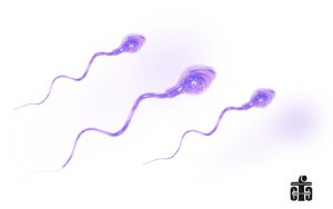
Al comparar ambos gametos el que sufre más lesiones en su material genético es el espermatozoide porque no cuenta con un sistema de reparación del ADN. Este motivo posiciona al espermatozoide como la célula sexual más comprometida en la fertilidad y por ello, en años recientes se denominó a la fragmentación del ADN espermático como principal causa molecular de infertilidad masculina que genera daños letales o subletales en los espermatozoides, alterando, en primer lugar, parámetros vinculados a su capacidad fecundante, y luego, comprometiendo a la descendencia (Agarwal et al., 2020; Esteves et al., 2021). Sosteniendo este nuevo paradigma en la infertilidad masculina, a la par de los avances sobre esta temática, se observó en múltiples casos clínicos una correlación entre dicho parámetro cuando supera el umbral de fragmentación (>20%) y los efectos negativos en los resultados reproductivos de las parejas, debido a la pobre calidad del embrión resultante en términos genéticos y morfológicos. Ante estos resultados, se concluyó que a mayor grado de fragmentación del ADN espermático aumentan los riesgos de producirse un fallo de implantación del embrión o abortos espontáneos recurrentes, disminuyendo la probabilidad de producir un embarazo a término (Agarwal et al., 2019).

No obstante, es importante mencionar que los resultados negativos vinculados a este parámetro seminal se reflejan tanto en la técnica de IIU como las de FIV e ICSI. En consecuencia, se intensificó la investigación en esta área, dando lugar al desarrollo de nuevas herramientas que procesen muestras seminales más eficientemente, incluyendo la integridad genética como parámetro central de selección (Rappa et al., 2016). A pesar de observarse resultados alentadores, especialmente con los dispositivos desarrollados a partir de microfluidos, muchas de las nuevas metodologías aún se encuentran bajo investigación para comprobar su eficacia y eficiencia en comparación con las técnicas convencionales originadas hace más de 30 años (Samuel et al., 2018).

La gran diferencia entre las técnicas rutinarias y las de vanguardia es que las últimas evitan pasos que dañan la integridad genética con la finalidad de recuperar los espermatozoides con poca o nula fragmentación de su material hereditario. Pero aún debe aumentar el volumen de estudios para garantizar su seguridad y ser avalados por la comunidad científica y clínica en la práctica. Sin duda alguna, el equipo de laboratorio coincide con otros especialistas en que el incremento de estudios sobre el impacto de herramientas innovadoras diseñadas principalmente desde el punto de vista genético aportará información relevante para aplicar en un futuro cercano herramientas terapéuticas más precisas, personalizadas e integrales acorde a las características de los pacientes afectados, logrando aumentar las tasas de embarazo.

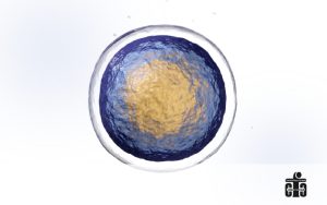
Mientras tanto, en condiciones naturales, la reparación de daños en el ADN del espermatozoide depende del ovocito que sí cuenta con un sistema de reparación que actúa tras ser fecundado por éste, cuando se complementan ambos genomas para constituir un cigoto diploide (2n). A pesar de contar con esta estrategia natural, la capacidad de reparación que tiene el ovocito va a depender del tipo de lesión que presente el ADN espermático y su porcentaje afectado, y la calidad del ovocito, un factor generalmente ligado a la edad de la mujer (Colaco y Sakas, 2018). Pero en este punto existe una limitación, ya que es más difícil evaluar el ovocito por ser una célula sexual compleja. Más aún son escasos los estudios sobre la fragmentación del ADN de ovocitos ya que la disponibilidad es acotada debido a que, a diferencia de los espermatozoides que se encuentran en constante producción, el gameto femenino se produce durante la vida fetal de la mujer y esta nace con entre 1 y 2 millones de ovocitos los cuales irá perdiendo a lo largo de su vida sin reponerlos. Esto hace que la reserva de ovocitos sea limitada y que su disponibilidad decrezca con la edad. A partir de ello, solo se han realizado pocos estudios sobre ovocitos donados para investigación por pacientes que obtuvieron un excedente de estos en sus punciones foliculares para tratamientos de FIV o ICSI. De hecho, es habitual en los laboratorios de reproducción humana realizar solo una evaluación morfológica de los ovocitos, ya que el usar técnicas más invasivas podría destruir las células reproductivas femeninas sin posibilidad de usarlas en el tratamiento. Por esta razón muchos trabajos publicados en el tema se basaron en resultados retrospectivos.

Distintos expertos coinciden, a partir de trabajos científicos, del diagnóstico morfológico de gametos y de los resultados clínicos, en que la calidad ovocitaria normalmente disminuye con el envejecimiento de la mujer, incluyendo su integridad genética, como consecuencia de factores estresantes que afectan al ovario y, por ende, reducen el número de ovocitos disponibles y su calidad, siendo además un efecto acumulativo en el tiempo (Winship et al., 2018; Ahmed et al., 2019). A raíz de ello se ha desarrollado la hipótesis de que la pérdida de calidad genética del gameto femenino desgasta la maquinaria de reparación del ADN y eventualmente, la capacidad de reparar los daños de su propio ADN y del ADN espermático. Como evidencia parcial de ello, en varios análisis de casos se ha visto que, si la fragmentación del ADN espermático es muy extensa, el embrión puede no desarrollarse o no implantarse en el útero, o puede ser abortado naturalmente en un estadio posterior (González-Marín et al., 2012). Estos resultados suelen observarse con mayor frecuencia en ovocitos de mujeres con edad materna avanzada, es decir, mayor a 40 años (Setti et al., 2021).

Los resultados mencionados anteriormente, que son producto de factores masculinos y femeninos combinados, hacen nuevamente que la fragmentación del ADN espermático sea un parámetro relevante al momento de evaluar cómo mejorar el éxito reproductivo y, por tanto, se intensifiquen los esfuerzos para hallar nuevas herramientas que propicien un escenario alentador para todos los pacientes que acuden a la ayuda profesional.

Por último, como la edad es un componente relacionado al envejecimiento reproductivo, también existen técnicas para preservar la fertilidad. La herramienta más importante para ello es la criopreservación, mediante la cual se conservan los gametos viables y se recurre a ellos en el momento que se desee concebir. Las técnicas de criopreservación más desarrolladas son el congelamiento lento y la vitrificación.

Una dificultad que presentan las técnicas de criopreservación es que, durante el procedimiento los gametos sufren un estrés fisiológico pudiendo generar en aquellas que sobreviven daños subcelulares e inducir la fragmentación del ADN (Ezzati et al., 2020; Albertini y Olsen, 2013), y luego, repercutir en la tasa de supervivencia de los gametos como también en la tasa de embarazo. Ante esta cuestión, estudios comparativos demostraron que la técnica de vitrificación es el método más eficiente y apropiado de criopreservación, porque la supervivencia de los gametos descongelados es mayor y además genera menor daño en el material genético (Iaizzo et al., 2016). Es por ello que numerosos laboratorios de reproducción asistida en todo el mundo la utilizan regularmente.

En síntesis, aún queda camino por recorrer en el campo de la medicina reproductiva para garantizar un embarazo exitoso y una descendencia sana. Con este fin, actualmente está teniendo especial interés el abordaje del factor genético desde la preconcepción hasta el futuro desarrollo del embrión en las técnicas de reproducción asistida humana para evitar anomalías genéticas y reducir los riesgos durante el embarazo. Particularmente, analizando los casos clínicos se halló que la fragmentación del ADN espermático tiene un impacto relevante en la fertilidad, siendo un parámetro inverso a los resultados reproductivos. Además, si ambos gametos tienen alteraciones genéticas severas existe la posibilidad de que sean incompatibles con el desarrollo embrionario y fetal normal, o presentar efectos negativos en las próximas generaciones, como la transmisión de enfermedades hereditarias. A partir de los hechos observados y junto al estado actual de los avances en tecnología y biología molecular, se está profundizando en este tema aún en exploración con el fin de desarrollar técnicas mejoradas de diagnóstico y tratamiento que asistan y con mayor precisión a cada paciente en su aspiración a concretar el nacimiento de un niño.

Numerosos grupos de investigación de todo el mundo están abocados al estudio de la alta fragmentación del ADN de los gametos, ya que se presenta como denominador común de varios tipos de efectos negativos en la fertilidad. Ahondar en este aspecto puede llegar a ser una solución definitiva para una población considerable de pacientes afectados por ella.


Importance of evaluating gamete DNA fragmentation in human assisted reproduction techniques

Human assisted reproductive techniques (ART) are biomedical tools that mimic the in vivo fertilization process in an artificial environment in vitro and are used to treat both male and female infertility that affects around 15% of couples in reproductive age worldwide, being more than 50 million people, according to the latest statistics from the World Health Organization (Babakhanzadeh et al., 2020).

This medical condition prevents conceiving naturally due to a set of multiple causes such as age, medical history, environmental factors and lifestyle, which often impact to some degree at the molecular level in the DNA of the gametes (Fainberg and Kashanian, 2019; Ahmed et al., 2020).

Specifically, unlike other causes of infertility such as metabolic ones, in those cases where infertility has a genetic factor, there are no pharmacological strategies to remedy it. Therefore, the alternative to this is to undergo some assisted fertility treatment to increase the chances of achieving pregnancy, and in turn, avoid genetic abnormalities in the offspring. Depending on the diagnosis determined by different professionals, the technique of intrauterine insemination (IUI), in vitro fertilization (IVF) or intracytoplasmic sperm injection (ICSI) is used. This last technique was developed in the 1990s, giving rise to a more specific treatment for male infertility, where a few spermatozoa are selected from a poor sample to be mechanically introduced into the oocytes (Rex et al., 2017). With this technique, the operator selects the fertilizing sperm that must always present good mobility because it is an indicator of vitality and genetic integrity.

Now, regardless of the technique to be used, the genomic integrity of the female and male gametes is of vital importance in this field, mainly because the embryo inherits a genetic load 50% maternal and 50% paternal, so that the greater is the parent’s DNA integrity the risks that lead to a failed treatment will be lower. In other words, when DNA presents structural damage, their integrity is lower, and therefore, the reproductive potential of the gamete decreases and, in the event of generating an embryo, compromise the normal development. In the opposite case, an adequate genetic integrity is favorable for the gamete since it will generate a viable embryo with greater chances of success in the treatment which will be achieved accompanied by the most appropriate technique for the circumstances.

When comparing both gametes, the one that suffers the most damage to its genetic material is the sperm because it does not have a DNA repair system. This reason positions the sperm as the most compromised sexual cell in fertility and for this reason in recent years sperm DNA fragmentation has been called the main molecular cause of male infertility that generates lethal or sub-lethal damage to the sperm, altering in first place parameters linked to its fertilizing capacity and then compromising the offspring (Agarwal et al., 2020; Esteves et al., 2021). Supporting this new paradigm in male infertility, along with advances on this subject, a correlation was observed in multiple clinical cases between said parameter when it exceeds the fragmentation threshold (>20%) and the negative effects on the reproductive results of couples, due to the poor quality of the resulting embryo in genetic and morphological terms. Given these results, it was concluded that a higher degree of sperm DNA fragmentation increases the risks of embryo implantation failure or recurrent spontaneous abortions, reducing the probability of producing a full-term pregnancy (Agarwal et al., 2019).

However, it’s important to mention that the negative results linked to this seminal parameter are reflected in both the IUI technique and those of IVF and ICSI. Consequently, the research in this area was intensified, leading to the development of new tools that process seminal samples more efficiently, including genetic integrity as a central selection parameter (Rappa et al., 2016).

Despite encouraging results, especially with devices developed from microfluidics, many of the new methodologies are still under investigation to verify their effectiveness and efficiency compared to conventional techniques originated more than 30 years ago (Samuel et al., 2018). The big difference between routine and cutting-edge techniques is that the new ones avoid steps that damage genetic integrity in order to recover spermatozoa with little or no fragmentation of their hereditary material, but the volume of studies must still be increased to guarantee their safety and be endorsed by the scientific and clinical community in practice. Without a doubt, the laboratory team agrees with other specialists that the increase in studies on the impact of innovative tools designed mainly from a genetic point of view will provide relevant information to apply more precise, personalized, and comprehensive therapeutic tools in the near future according to the characteristics of the affected patients, managing to increase pregnancy rates.

Meanwhile, under natural conditions, the repair of sperm DNA damage depends on the oocyte, which does have a repair system that acts after being fertilized by it, when both genomes complement each other to form a diploid zygote (2n). Despite having this natural strategy, the repair capacity of the oocyte will depend on the type of damage that the sperm DNA presents and its affected percentage, and the quality of the oocyte, a factor generally linked to the age of the woman (Colaco and Sakas, 2018). But at this point there is a limitation since it’s more difficult to evaluate the oocyte because it’s a complex sexual cell. Furthermore, there are few studies on the DNA fragmentation of oocytes since availability is limited because unlike spermatozoa which are in constant production, the female gamete is produced during the woman’s fetal life, and she is born with between 1 and 2 million oocytes which she will lose throughout her life without replacing them. This means that the oocyte reserve is limited, and its availability decreases with age.

From this only few studies have been carried out on oocytes donated for research by patients who obtained a surplus of them in their follicular punctures for IVF or ICSI treatments. In fact, it’s common in human reproduction laboratories to perform only a morphological evaluation of the oocytes, since using more invasive techniques could destroy the female reproductive cells without the possibility of using them in their treatment. That is why many published works on the subject were based on retrospective results.

Based on scientific studies, the morphological diagnosis of this gamete and clinical results, different experts agree that oocyte quality normally decreases with women aging, including their genetic integrity, as a consequence of stressful factors that affect the ovary and therefore, they reduce the number of available oocytes and their quality, also being a cumulative effect over time (Winship et al., 2018; Ahmed et al., 2019). As a result of this, the hypothesis has been developed that the loss of genetic quality of the female gamete wears out the DNA repair machinery and eventually, the ability to repair damage to its own DNA and to sperm DNA. As partial evidence of this, several case studies have shown that if the sperm DNA fragmentation is very extensive, the embryo may not develop or implant in the uterus, or it may be aborted naturally at a later stage (González-Marín et al., 2012). These results are usually observed more frequently in oocytes from women with advanced maternal age, older than 40 years (Setti et al., 2021).

The results mentioned above, which are the product of combined male and female factors, once again make sperm DNA fragmentation a relevant parameter when evaluating how to improve reproductive success, and therefore efforts are intensified to find new tools that promote an encouraging scenario for all patients who seek professional help.

Lastly, as age is a component related to reproductive aging, there are also techniques to preserve fertility. The most important tool for this is cryopreservation, by which viable gametes are preserved and used at the time you want to conceive. The most developed cryopreservation techniques are slow freezing and vitrification.

One difficulty with cryopreservation techniques is that during the procedure the gametes suffer physiological stress, which can generate subcellular damage in those that survive and induce DNA fragmentation (Ezzati et al., 2020; Albertini and Olsen, 2013), and then affect the survival rate of gametes as well as the pregnancy rate. Given this situation, comparative studies have shown that the vitrification technique is the most efficient and appropriate method of cryopreservation, because the survival of thawed gametes is greater and it also generates less damage to the genetic material (Iaizzo et al., 2016). That is why many assisted reproduction laboratories around the world use it regularly.

In synthesis, there is still a long way to go in the field of reproductive medicine to ensure a successful pregnancy and healthy offspring. To this end, the approach to the genetic factor from preconception to the future development of the embryo in human assisted reproduction techniques is currently of special interest in order to avoid genetic anomalies and reduce risks during pregnancy. Analyzing clinical cases, it was found that sperm DNA fragmentation has a relevant impact on fertility, being an inverse parameter to reproductive results. In addition, if both gametes have severe genetic alterations, there exist the possibility that they are incompatible with normal embryonic and fetal development, or have negative effects on future generations, such as the transmission of hereditary diseases. Based on the observed facts and together with the current state of advances in technology and molecular biology, this subject is still being explored in depth to develop improved diagnostic and treatment techniques that assist each patient with greater precision in his aspiration to realize the birth of a child.

Numerous research groups around the world are devoted to the study of the high fragmentation of gamete DNA, since it is presented as a common denominator of several types of negative effects on fertility. Delving into this aspect can become a definitive solution for a considerable population of patients affected by it.


Bibliografía

Agarwal A, et al. Sperm DNA damage and its impact on male reproductive health: a critical review for clinicians, reproductive professionals and researchers. Expert Rev Mol Diagn. 2019 Jun;19(6):443-457. doi: 10.1080/14737159.2019.1614916

Agarwal A, et al. Sperm DNA Fragmentation: A New Guideline for Clinicians. World J Mens Health. 2020 Oct;38(4):412-471. doi: 10.5534/wjmh.200128

Ahmed TA, et al. Oocyte Aging: The Role of Cellular and Environmental Factors and Impact on Female Fertility. Adv Exp Med Biol. 2020;1247:109-123. doi: 10.1007/5584_2019_456

Albertini DF, Olsen R. Effects of fertility preservation on oocyte genomic integrity. Adv Exp Med Biol. 2013;761:19-27. doi: 10.1007/978-1-4614-8214-7_3. PMID: 24097379.

Babakhanzadeh E, et al. Some of the Factors Involved in Male Infertility: A Prospective Review. Int J Gen Med. 2020 Feb 5;13:29-41. doi: 10.2147/IJGM.S241099

Colaco S, Sakkas D. Paternal factors contributing to embryo quality. J Assist Reprod Genet. 2018 Nov;35(11):1953-1968. doi: 10.1007/s10815-018-1304-4.

Esteves SC, et al. Sperm DNA fragmentation testing: Summary evidence and clinical practice recommendations. Andrologia. 2021 Mar;53(2):e13874. doi: 10.1111/and.13874

Ezzati M, et al. Influence of cryopreservation on structure and function of mammalian spermatozoa: an overview. Cell Tissue Bank. 2020 Mar;21(1):1-15. doi: 10.1007/s10561-019-09797-0

Fainberg J, Kashanian JA. Recent advances in understanding and managing male infertility. F1000Res. 2019 May 16;8:F1000 Faculty Rev-670. doi: 10.12688/f1000research.17076.1

González-Marín C, et al. Types, causes, detection and repair of DNA fragmentation in animal and human sperm cells. Int J Mol Sci. 2012 Oct 31;13(11):14026-52. doi: 10.3390/ijms131114026

Iaizzo, et al. Efecto de la criopreservación sobre la integridad del ADN del oocito. Reproducción. 2016 Marzo 31(1):3-10. URL: http://www.samer.org.ar/revista/numeros/2016/17-%20G%20MARTINEZ%202.pdf

Rappa KL, et al. Sperm processing for advanced reproductive technologies: Where are we today? Biotechnol Adv. 2016 Sep-Oct;34(5):578-587. doi: 10.1016/j.biotechadv.2016.01.007

Rex AS, et al. DNA fragmentation in spermatozoa: a historical review. Andrology. 2017 Jul;5(4):622-630. doi: 10.1111/andr.12381

Samuel R, et al. Microfluidic-based sperm sorting & analysis for treatment of male infertility. Transl Androl Urol. 2018 Jul;7(Suppl 3):S336-S347. doi: 10.21037/tau.2018.05.08

Setti AS, et al. Oocyte ability to repair sperm DNA fragmentation: the impact of maternal age on intracytoplasmic sperm injection outcomes. Fertil Steril. 2021 Jul;116(1):123-129. doi: 10.1016/j.fertnstert.2020.10.045

Winship AL, et al. The importance of DNA repair for maintaining oocyte quality in response to anti-cancer treatments, environmental toxins and maternal ageing. Hum Reprod Update. 2018 Mar 1;24(2):119-134. doi: 10.1093/humupd/dmy002


Historial de publicación

16 febrero 2022

Palabras clave

Infertilidad, fragmentación del ADN espermático, técnicas de reproducción asistida


Abrir chat